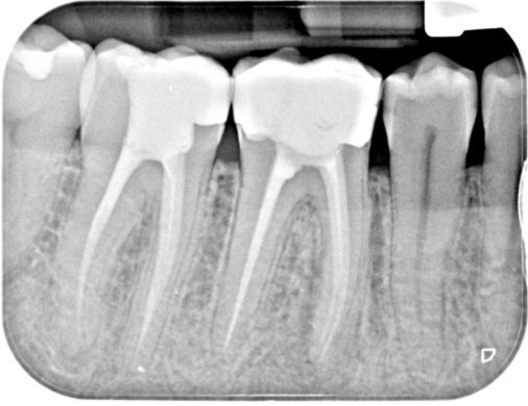
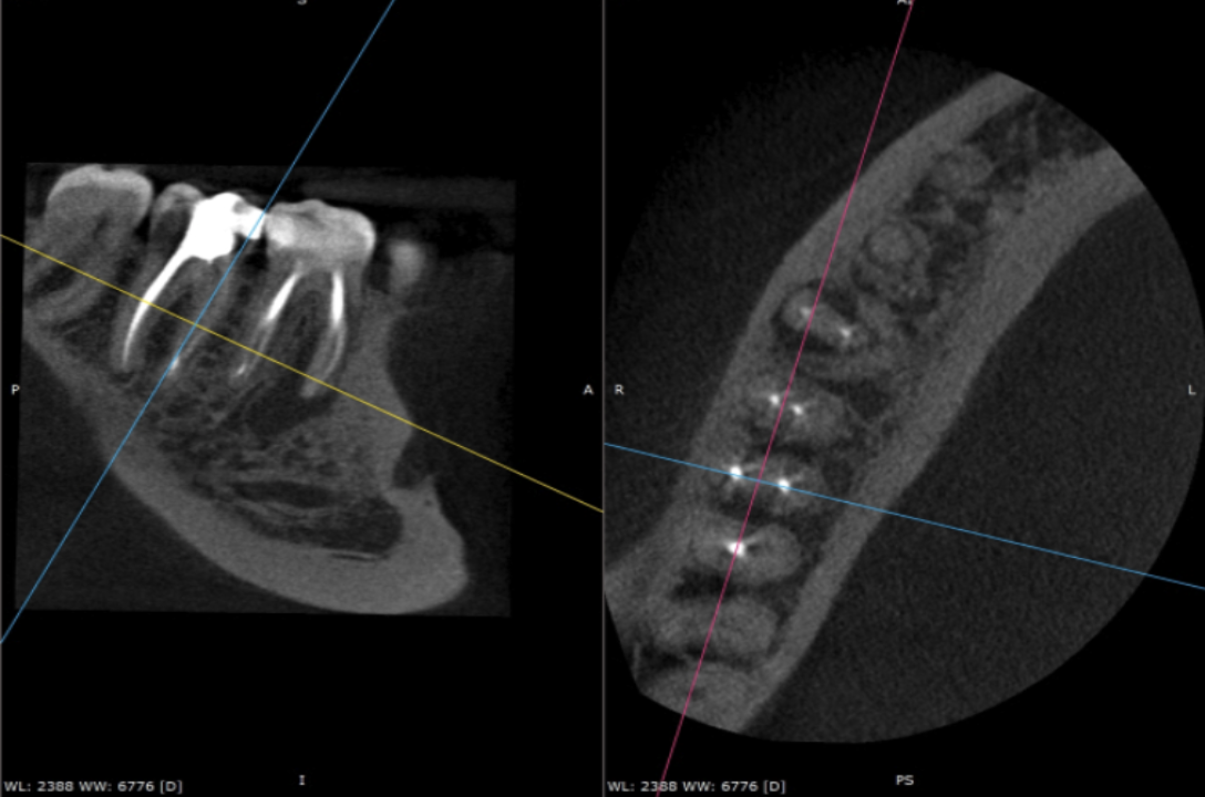
This was an interesting case from a diagnostic perspective. At the consultation appointment, the patient reported previous episodes of poorly localised pain from the LRQ and a recent episode of swelling in this area (not present at the consultation). The RD had managed this with antibiotics. IOPA review indicated that the LR6 and LR7 had been previously root treated (well!), but it was unclear which tooth was problematic. The patient reported that both root treatments were performed approximately 7-8 years ago. As both teeth had large composites in situ, a loss of coronal seal seemed like a possible cause of failure for either root treatment. In an attempt to clarify things and to help reach a diagnosis, we took a small field of view high-resolution CBCT scan of the area (see above). I requested a consultant radiologist’s report on the CBCT scan from JM Radiology with the key findings listed below:
- A significant PARL around the mesial root of the LR6 that extended halfway up the proximal surface.
- There were 4 root canals, 2 mesial and 2 distal. The 2 mesial canals had separate exit portals, and the 2 distal canals exited through a single exit portal (extremely useful information to have when you are planning a re-treatment case)
- No unfilled canal space (or was there!)
- Could not discount a fracture, therefore the patient had to be consented for this.
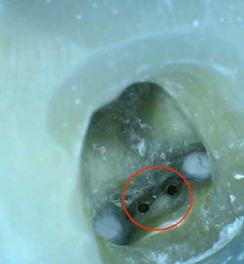
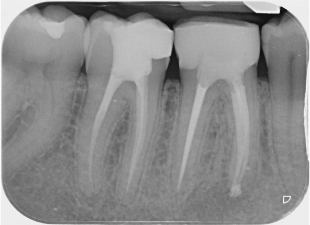
A diagnosis of failed root treatment with a recent acute apical abscess was made for the LR6. Once this tooth was accessed utilising an operating microscope, the most likely cause of failure was identified and can be seen in the picture below. There were 2 unfilled middle mesial canals not identified by the scan and this seemed to explain the pattern of inflammatory change seen around the mesial root. As helpful as CBCT imaging can be in endodontics, this case highlights that CBCT analysis of root-treated teeth does have its limitations. Scatter artefact can often make it quite difficult to see fine detail in such cases, so it’s useful to bear this in mind. This case was retreated using Reciproc Blue R25 and involved copious amounts of irrigation and activation. It was then obturated using hydraulic condensation and Totalfill BC sealer Hiflow. It was then sent back to the RD for cuspal coverage. I have suggested getting a follow-up CBCT scan in 12 months’ time to assess healing and also to review the small radiolucency at the distal apex of the LR7. I will update this case study once I have the review imaging.
Photo of middle mesial canals and post-op IOPA
With the option for self-referrals and referrals by your dentist, Modern Endodontics by Dr Adam Watt makes it easy for root canal patients based in Glasgow and further afield to access advanced endodontic care.
If you're a patient looking to self-refer for an endodontic treatment with Dr Adam Watt, please contact the Treatment Coordinator at Broomhill Dental, Glasgow.